What’s Your Diagnosis? Challenge Answer April – June 2012
Special thanks to Dr. Brian Rois-Mendez of Acacia Animal Hospital for referring this case to us.
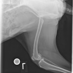
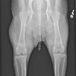
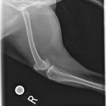
This newsletter’s challenge is an 11-year-old M/C Australian Shepherd presenting chronic lameness on the left hind limb, with pain isolating to the left stifle. Medial buttress sign to the left stifle. Three views of the pelvis and stifles are available for interpretation.
WYD Answer:
Hips: The coxofemoral joints appear normal. There is a large fat dense mass associated with the caudal aspect of the right thigh. Mild atrophy of the left thigh musculature is present.
Left stifle: There is moderate periarticular osteophyte formation and moderate intracapsular soft tissue swelling present. On the lateral projection there are multiple small osseous bodies overlying the cranial aspect of the stifle joint.
Right stifle: Changes to the right stifle are similar to those on the left less the overlying osseous bodies.
Conclusion:
1. Bilateral stifle joint effusion and moderate DJD consistent with chronic ligamentous instability. Bilateral cruciate disease is most likely.
2. Right caudal thigh lipoma. Distinction between infiltrative and intermuscular lipoma should be made prior to surgery. Consider a CT study of the right thigh.
3. Mild atrophy to the left thigh musculature consistent with historical lameness.
Many of you correctly identified the stifle disease, but did not mention the lipoma. These lipomas, or “fatty masses” can be trouble because they can infiltrate the muscle. When muscle infiltration occurs, they are called infiltrative lipomas.
CT is the quickest and one of the best ways to evaluate these lipomas.
A comprehensive analysis of infiltrative lipomas is found in the article “Computed Tomographic Imaging of Infiltrative Lipoma in 22 Dogs” in the 2001 May-June issue of the journal Veterinary Radiology & Ultrasound.


